The Benefits of Elastography in Ultrasound
The Rise of Ultrasound Elastography
Based on calls and requests from our customers, we’re finding that elastography is quickly becoming one of the more popular emerging technologies in ultrasound.
And based on calls from manufacturers, they’re on it. We keep getting excited calls from them saying “We Have Elastography on another model!”
Then, of course, the their next sentence is “Sooooo…. How many more can you sell next month?”
Well, they don’t ALL say that… or maybe I’m just feeling that vibe.
Anyway, the point is that Elastography is making its way to the mainstream.
This rise in popularity is the result of a few factors: price, availability, and expanded clinical applications.
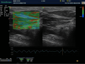
Elastography is traditionally known for its use with breast ultrasound in helping identify malignant vs benign breast lesions. These days, it is commonly found as a routine part of a breast examination because it typically results in a reduced number of biopsies.
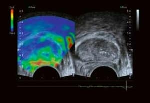
More recently, however, elastography has become more affordable and is found on many ultrasound machines. Because of this, clinicians are finding new ways to use ultrasound elastography, such as identifying liver, thyroid, and prostate cancers. Now, elastography has found its way into the MSK field.
What is Elastography?
In short, elastography maps the “elastic properties” (stiffness) in tissue. This is important because diseased tissue is most often stiffer than normal tissue. Using various methods, the ultrasound can display or quantify various levels of stiffness in tissue.
Think visual palpation.
There are two common elastography techniques found on ultrasound machines: Compression (strain) and Shear Wave:
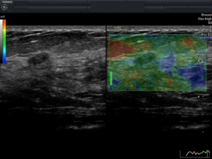
Compression, or “strain“, is the most affordable and commonly found ultrasound elastography option. It is performed by manually compressing tissue with an ultrasound transducer, and the ultrasound color-codes the various levels of stiffness. The drawback to this method is that it doesn’t provide quantitative results, as the amount of compression applied cannot be measured by the ultrasound. Still, it’s found to be very helpful and is considered the “new palpation” method to identifying diseased tissue.
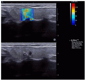
Shear Wave is the more advanced method. It’s considered more beneficial because it provides quantitative results to the elasticity of tissue, which can be especially helpful when comparing tissue stiffness over time. Shear Wave Elastography is a more complex and advanced method because it uses a transducer that can perform a specific task when sending and receiving an ultrasonic signal, which allows it to measure a specific amount of stiffness. At this time, it is a much more expensive option and not necessarily advantageous in situations where the standard manual compression method will suffice to identify tissue stiffness.
Which Ultrasounds have Elastography?
For the common compression technique, a largely increasing number of ultrasound machines have an elastography option. This includes many affordable mid-range ultrasound systems, including newer systems from Chison and SonoScape. However, you typically will not find it on lower-end color systems.
A small percentage of ultrasound machines have Shear Wave or similar technologies. It is a more advanced technology, and thereby is currently found on only high-end, advanced, expensive ultrasound machines from manufacturers such as Philips, GE, Toshiba, and Siemens.
All systems are good for basic qualitative analysis, identifying a nodule and determining its stiffness compared to the surrounding tissue. For most of our customers, the standard strain imaging provides them what they need.
Interested in getting more information or purchasing an ultrasound for elastography? Call Probo Medical today at 866-513-8322 to talk to one of our sales experts about your ultrasound needs.
About the Autor
Brian Gill is Probo Medical’s Vice President of Marketing. He has more than 20 years of experience in the ultrasound industry. From sales to service to customer support, he has done everything from circuit board repair and on-site service to networking and PACS, to training clinicians on ultrasound equipment. Through the years, Brian has trained more than 500 clinicians on over 100 different ultrasound machines. Currently, Brian is known as the industry expert in evaluating ultrasounds and training users on all makes and models of ultrasound equipment, this includes consulting with manufacturers with equipment evaluations during all stages of product development.

